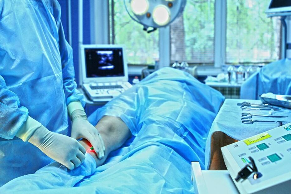
The main treatment for varicose veins (VV) remains surgery. The purpose of surgery is to eliminate symptoms of the disease (including cosmetic defects) and prevent the progression of saphenous varicose vein transformation. Nowadays, none of the existing surgical methods is suitable by itself for treatment of all pathogenesis; therefore, the necessity of combining them becomes obvious. Various combinations of certain surgeries mainly depend on the severity of the disease in the venous system of the lower limbs.Indications for surgery are the presence of blood reflux from deep veins to superficial veins in patients with grades C2-C6. Combining operations can include the following steps:
ligation and crossing of the GSV and/or SVC with the estuaries of all tributaries (cross resection);
Remove GSV and/or SSV trunks;
Removal of varicose tributaries of GSV and SSV;
Incompetent crossing of perforating veins.
This operating range has been developed over decades of scientific and practical research.Cross resection of the great saphenous vein. The best way to connect the GSV is through the inguinal fold. Due to the high location of the residual pathological stump of GSV and the postoperative scar, the suprainguinal approach has certain advantages only in patients with recurrent disease. The GSV must be ligated strictly to the wall of the femoral vein; all estuarine tributaries, including the upper tributaries (superficial epigastric vein), must be ligated. No suturing of the oval window or subcutaneous tissue is required after GSV transection.Resection of the great saphenous vein trunk. When determining the extent of GSV detachment, it must be taken into account that in the vast majority of cases (80-90%), only backflow along the GSV from the oral cavity to the upper third of the leg is recorded. Compared with removal of the GSV from the mouth to the upper third of the leg (short dissection), removal of the GSV along its entire length (full dissection) significantly increased the incidence of saphenous nerve injury - 39% versus 6. 5%, respectively. At the same time, there was no significant difference in the frequency of varicose vein recurrence. Remaining portion of vein could be used for reconstructive vascular surgery in the futureIn this regard, the basis for intervention in the GSV basin should be short stripping. Resection of the entire trunk was allowed only if it was reliably confirmed that the trunk was nonfunctional and significantly dilated (more than 6 mm in horizontal position).When choosing a safe resection, intussusception techniques (including PIN stripping) or cryophlebectomy should be given priority. Although detailed studies of these methods are still ongoing, their advantages (less invasive) compared with the classic Babcock technique are unquestionable. However, the Babcock method is effective and can be used clinically, but it is recommended to use small diameter olives. When choosing the direction of vein resection, priority should be given to traction from top to bottom, that is, retrograde, except for cryophlebectomy, which involves antegrade resection of the vein.Cross resection of the small saphenous vein. The structure of the terminal portion of the small saphenous vein is highly variable. Typically, the SVC joins the popliteal vein a few centimeters above the knee flexion line. In this regard, the approach to superior vena cava cross resection must be shifted proximally, taking into account the positioning of the saphenopopliteal anastomosis (the positioning of the anastomosis should be clarified using preoperative ultrasound scanning).The trunk of the lesser saphenous vein is removed. As with GSV, the vein should be excised only if reflux is established. Reflux along the superior vena cava in the lower third of the leg is very rare. The invagination method should also be used. SVC cryophlebectomy offers no advantages over these techniques.One comment. Interventions on the small saphenous vein (transection and trunk resection) should be performed with the patient in the prone position.Thermal occlusion of the major saphenous vein. Modern endovascular techniques—lasers and radiofrequency—can eliminate brainstem reflux and, therefore, can be called alternatives to transection and dissection in terms of their functional effectiveness. Thermal obliteration is associated with significantly lower morbidity than stylovein resection and significantly greater cosmetic results. Laser and radiofrequency ablation eliminate the need for orifice ligation (GSV and SSV). Simultaneous transection virtually eliminates the benefits of thermal obliteration and increases the cost of treatment.Endovascular laser and radiofrequency ablation have limitations in use, are associated with specific complications, are expensive, and require mandatory intraoperative ultrasound control. The technique has low reproducibility and therefore can only be performed by experienced experts. The long-term results of use in widespread clinical practice remain unknown. In this regard, thermal ablation methods require further research and cannot completely replace traditional surgical intervention methods for varicose veins. Remove varicose veins. When eliminating variceal tributaries of superficial trunks, their removal by skin puncture using venous excision instruments should be preferred. All other surgical methods are more traumatic and result in poorer cosmetic results. With the patient's consent, some varicose veins can be left in place and subsequently eliminated using sclerotherapy.Anatomy of perforating veins. The main controversial issue in this subsection is the determination of indications for intervention, since the role of perforators in the development of chronic venous disease and its complications needs to be clarified. The inconsistency among numerous studies in this field is related to the lack of clear criteria for determining perforating vein insufficiency. Many authors generally question the fact that incompetent perforating veins may have an independent significance in the development of CVD and be the source of pathological reflux from the deep to the superficial venous system. The primary function of varicose veins is vertical drainage through the great saphenous vein, and failure of the perforators is associated with increased load on the perforators to drain returning blood from the superficial to the deep venous system. As a result, they increase in diameter and have bidirectional blood flow (mainly into deep veins), which mainly depends on the severity of vertical reflux. It should be noted that bidirectional blood flow across perforators has also been observed in healthy individuals without evidence of CVD. The number of incompetent perforating veins correlates directly with CEAP clinical classification. These data are partially corroborated by studies in which a significant proportion of the perforators became solvent after intervention in the superficial venous system and elimination of reflux.However, in patients with nutritional diseases, 25. 5% to 40% of perforators remain incompetent and their further impact on the course of the disease is unknown. Clearly, for C4-C6 varicose veins, the possibility of restoring normal hemodynamics in perforating veins after elimination of vertical reflux is limited. As a result of long-term exposure to pathological reflux from subcutaneous and/or deep veins, certain parts of these vessels undergo irreversible changes, and the reflux of blood through them has pathological significance.Therefore, today we can discuss the mandatory careful ligation of incompetent perforating veins only in patients with varicose veins with nutritional disorders (categories C4-C6). In clinical categories C2-C3, the decision to ligate the perforator must be made individually by the surgeon based on the clinical situation and instrumental examination data. In this case, dissection should be performed only after its failure has been reliably confirmed.If the localization of the dystrophic disorder precludes the possibility of direct percutaneous access to an incompetent perforating vein, the procedure of choice is endoscopic perforating vein dissection (ESDPV). Numerous studies have shown its undeniable advantages over the previously widely used open subfascial perforator ligation (Linton procedure). The wound complication rate for ESDPV is 6-7% compared with 53% for open surgery. At the same time, the healing time, venous hemodynamic parameters, and recurrence frequency of trophic ulcers were comparable.One comment. Numerous studies have shown that ESDPV can have a positive impact on the course of chronic venous disease, particularly with regard to nutritional disorders. However, it is unclear which of the observed effects are due to anatomy and which are due to the concurrent saphenous vein surgery performed in most patients. However, patients with C4-C6 did not undergo perforating venous intervention, only phlebectomy, and long-term results are lacking, so we cannot yet draw final conclusions about the use of certain surgical treatments.Despite the contradictions, most researchers still consider it necessary to combine traditional superficial venous intervention with ESDPV to treat patients with nutritional diseases and open trophic ulcers in the context of varicose veins. Ulcer recurrence rates after combined phlebectomy and ESDPV range from 4% to 18% (follow-up period 5-9 years). In this case, approximately 90% of patients will fully recover within the first 10 months.When other minimally invasive techniques are used to eliminate perforator veins, such as microfoam sclerosing occlusion, endoluminal laser occlusion, etc. , good results are also achieved. However, the likelihood of success in its use depends directly on the qualifications and experience of the physician, so widespread use is not currently recommended.ESDPV should not be used in patients with clinical classification C2-C3, as perforator reflux can be successfully eliminated through small incisions (maximum 1 cm) or even skin puncture using small phlebotomy instruments. Correction of deep vein valves. Currently, there are more questions than answers in this area of surgical phlebology. This is due to the contradictions in the significance of deep venous reflux and its impact on the course of CVI, determination of correction indications, and evaluation of treatment effects. Failure of various segments of the deep venous system of the lower extremities can lead to various hemodynamic disorders, which is an important factor to consider when selecting treatment methods. Numerous studies have shown that return flow through the femoral vein does not play any important role. At the same time, damage to the deep veins in the legs can lead to irreversible changes in muscle venous pump function and severe CVI. It is difficult to assess the positive effect of correcting venous reflux in the deep veins themselves, since these interventions are in most cases performed in conjunction with superficial and perforator vein surgeries. Elimination of reflux through the femoral vein alone either does not affect venous hemodynamics at all or results in only minor temporary changes in certain parameters. On the other hand, only elimination of reflux along the GSV in the varicose veins combined with femoral venous insufficiency can restore valvular function in this venous segment.Surgical approaches to treating primary deep venous reflux can be divided into two groups. The first involves phlebotomies, including internal valvuloplasty, transposition, autografting, creation of new valves, and use of cryopreserved allografts. The second group did not require phlebotomy and included extravascular intervention, external valvuloplasty (transmural or transcommissural), angioscope-assisted extravascular valvuloplasty, and percutaneous installation of corrective devices.The question of deep venous valve correction should be raised only in patients with recurrent or non-healing trophic ulcers (grade C6), mainly recurrent trophic ulcers and grade 3-4 deep venous reflux (up to grade C6). Knee joint) according to Kistner classification. Surgery can be performed for severe edema and C4b in young people who do not want to wear compression stockings for the rest of their lives if conservative treatment fails. Surgical decisions should be made based on clinical status rather than data from specialized studies, as symptoms may not correlate with laboratory parameters. Surgery to correct deep vein valves can only be performed in specialized centers with experience in this type of intervention.
Surgical treatment of post-thrombotic disease
The surgical treatment effect of PTB patients is significantly worse than that of varicose vein patients. Therefore, within the first 3 years after ESDPV, the recurrence rate of trophic ulcers reaches 60%. Many studies have not confirmed the effectiveness of venous perforator intervention in such patients.Patients should be informed that surgical treatment of PTB has a high risk of failure.
Intervention of the subcutaneous venous system
In many patients, the saphenous vein plays a collateral function in PTB, and removal of the saphenous vein may worsen the disease. Therefore, phlebectomy (as well as laser or radiofrequency obliteration) cannot be performed as a routine procedure for PTB. The decision on the need and possibility of excision of one or another volume of subcutaneous veins should be made based on a thorough analysis of clinical and anamnestic information, results of instrumental diagnostic tests (ultrasound, radionuclide).
Deep vein valve correction
In most cases, postthrombotic damage to the valve device is not amenable to direct surgical correction. Dozens of surgical options to create valves in deep veins to treat PTB have not yet moved beyond clinical trials.
Bypassing interventions
In the second half of the last century, two shunt interventions were proposed for deep venous occlusion, one of which aimed at diverting blood from the popliteal vein to the GSV in case of femoral occlusion (Warren-Tyre method), and the other -In the case of iliac vein occlusion, from the femoral vein to the other (healthy) limb (Palma-Esperon method). Only the second approach has shown clinical effectiveness. Not only is this procedure effective, it is the only method available today to create additional pathways for venous blood outflow and can be recommended for widespread clinical use. Autologous femoral-femoral cross vein shunts are characterized by lower thrombogenicity and better patency than artificial shunts. However, existing studies on this issue include a small number of patients with unclear clinical and venographic follow-up periods.Indications for femoral bypass surgery are unilateral iliac vein occlusion. The prerequisite is that there is no obstruction to the venous outflow of the contralateral limb. Furthermore, despite several (3–5) years of adequate conservative treatment, functional indications for surgery arise only when CVI has steadily progressed (to clinical categories C4–C6).
Vein grafting and transposition
Grafting of valve-containing vein segments has shown good success in the months following surgery. Usually the superficial vein of the upper limb is transplanted to the position of the femoral vein. The limitation of this method is due to differences in vein diameter. This intervention lacks pathophysiological justification: the hemodynamic conditions of the upper and lower limbs are significantly different, so the transplanted venous segment dilates with the development of reflux. Furthermore, replacing a severely damaged 1-2-3 valve in the deep venous system cannot compensate for the damaged venous outflow.Methods of recanalizing venous transposition "under the protection" of an intact vascular valve, of which from a technical point of view the most likely is transposition of the superficial femoral vein to the deep femoral vein, but are not recommended for widespread clinical use due to their complexityand the rarity of optimal implementation conditions in practice. Due to the small number of observations and lack of long-term results, we cannot draw any conclusions.
Endovascular intervention for deep vein stenosis and occlusion
Deep vein occlusion or stenosis is the primary cause of CVI symptoms in approximately one-third of patients with PVT. In the structure of trophic ulcers, 1% to 6% of patients have this pathology. Occlusion was accompanied by reflux in 17% of cases. Notably, this combination was accompanied by the highest levels of venous hypertension and the most severe CVI manifestations compared with reflux or occlusion alone. Proximal occlusion, especially of the iliac veins, is more likely to result in CVI than distal segmental involvement. Only 20-30% of iliac veins are completely recanalized due to iliofemoral thrombosis; in other cases, residual occlusion and the formation of more or less pronounced collateral circulation are observed. The primary goal of intervention is to remove or eliminate the occlusion or to provide additional pathways for venous outflow.Indications. Unfortunately, there are no reliable criteria for "severe stenosis" of the venous system. This is a major obstacle to determining indications for treatment and interpreting its results. X-ray contrast venography is a standard method for visualizing the venous bed and can determine the presence of areas of occlusion, stenosis, and collateral circulation. Intravascular ultrasonography (IVUS) is superior to venography in assessing the morphological characteristics and extent of iliac vein stenosis. Iliocaval segment occlusion and associated abnormalities can be diagnosed by MRI and spiral CT venography.Femoral iliac stenting. The introduction of percutaneous iliac vein balloon dilatation and stenting into clinical practice has significantly expanded treatment options. This is due to their high efficiency (segmental patency is restored in 50-100% of cases), low complication rates, and no deaths. Among the factors that cause thrombosis or restenosis in the stent area in patients with post-thrombophlebitis, the main factors are thrombophilia and excessive stent length. If these factors are present, restenosis rates after 24 months are as high as 60%; in the absence of these factors, no stenosis occurs. The healing rate of trophic ulcers after balloon dilation and iliac vein stent placement was 68%; 62% of cases did not relapse 2 years after intervention. The severity of swelling and pain was significantly reduced. The proportion of limbs experiencing swelling decreased from 88% to 53%, and the proportion of limbs experiencing pain decreased from 93% to 29%. Analysis of questionnaires in patients after venous stenting showed significant improvements in all major aspects of quality of life.Published venous stent studies often suffer from the same shortcomings as reports of open surgical interventions (small patient numbers, lack of long-term results, failure to group patients according to occlusion etiology, acute or chronic pathology, etc. ). Venous stenting has emerged relatively recently, so patient observation time has been limited. Because the long-term results of this procedure are unknown, continued monitoring is needed for several years to evaluate its effectiveness and safety.
Surgical treatment of venous hyperplasia
There is currently no effective method to cure hemodynamics in patients with venous hyperplasia. Surgery is needed when the saphenous vein is dilated and thin or when there is a risk of bleeding from a trophic ulcer. In these cases, venous mass resection is performed to reduce local venous stagnation.CVD surgery can be performed in vascular surgery or general surgery by specialists trained in phlebology. Certain types of interventions (reconstructive: valvuloplasty, bypass surgery, transposition, transplantation) can only be performed in specialized centers according to strict indications.